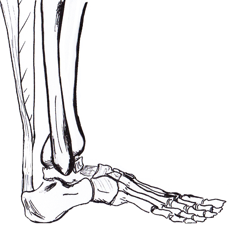
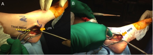
anatomy

Sesamoids

Anatomy


3 Sesamoids may be present in great toe

- 2 almost always present on plantar aspect of MTPJ

- 1 may be present on plantar aspect of IPJ


MTPJ sesamoids most important

- embedded in FHB tendons

- held together by intersesamoid ligament & plantar plate

- each side of crista / inter-sesamoid ridge

- articulate with plantar facets of 1st MT


Tibial usually larger than fibula

Achilles Tendonopathy

Definition


Inflammation of achilles tendon; insertional or noninsertional


Spectrum


Tendonitis / Tendonosis / Rupture


Anatomy


Triceps surae

- medial and lateral gastrocnemius

- soleus

- surrounded by paratenon which allows gliding and supplies nutrition


Inserts middle 1/3 calcaneal tuberosity

- 2 x 2 cm area

- 90o rotation distally


Retrocalcaneal bursa (x2)

Achilles Tendon Rupture

AnatomyAchilles tendon anatomy


Gastrocnemius tendon 10-25 cm long

- soleus 3-10 cm

- inserts superior calcaneal tuberosity

- fibres spiral 90°

- fibres that lie medially in proximal portion become posterior distally

- allows elastic recoil & energy storage


Plantaris present in 90% population

- medial to T Achilles


Poor blood supply midportion

- mesotenal vessels

Plantar Fasciitis

Definition


Pain at attachment of thickened central part of plantar aponeurosis to Medial Calcaneal Tuberosity


Anatomy Plantar Fascia


Origin 

- medial calcaneal tuberosity


Inserts 

- 5 bands superfical & deep layers


Superficial

- insert transverse MT ligament & skin


Deep 

- flexor sheath, volar plate & periosteum of P1


Tendonitis / Tendonopathy

Anatomy


Lateral compartment of leg

- run through retromalleolar groove

- pass superior and inferior to peroneal tubercle

- covered by inferior peroneal retinaculum


Peroneus longus

- origin lateral condyle of tibia and head fibula

- tendon PL superficial and inferior to brevis in retromalleolar groove

- runs in cuboid groove 

- insert plantar surface base of 1st MT and lateral aspect medial cuneiform

Dislocation

Definition


Anterior displacement of peroneal tendons out of peroneal groove


Epidemiology


Most common in young adults


Acute injury often missed 


Aetiology


Congenital


3 % neonates

- resolves spontaneously


Traumatic  


Occurs following sporting activities

Management

Definition


Complete Lesion 

- bulbocavernosus reflex present 

- no cord function below lesion

- very poor prognosis for recovery


Incomplete Lesion

- bulbocavernosus reflex present

- some cord function below lesion

- good prognosis for recovery


Anatomy


Vertebral Canal narrowest at T8/9

- Also area of vascular watershed


Cauda Equina

Definition


Compression of some or all of the nerve roots in the cauda equina

- bladder dysfunction

- bowel dysfunction

- saddle anaesthesia

- variable motor and sensory loss


Aetiology


HNP

- most common


Epidural haematoma

- post surgical

- spinals and anticoagulation


Cervical Radiculopathy

Definition


Clinical diagnosis

- based on a sclerotomal distribution of motor &/or sensory symptoms or signs


Caused by impingement of exiting nerve roots

- HNP

- zygo-apophyseal / facet joint hypertrophy

- neuro-central joint hypertrophy


May be acute or chronic


Epidemiology


M>F


Peak age 50-54


C7 > C6